This Case Study spotlight features insights from Dr. Kristopher Brand! Drawing from his extensive clinical experience, Dr. Brand offers a behind-the-scenes look at his diagnostic process, highlighting key imaging findings:
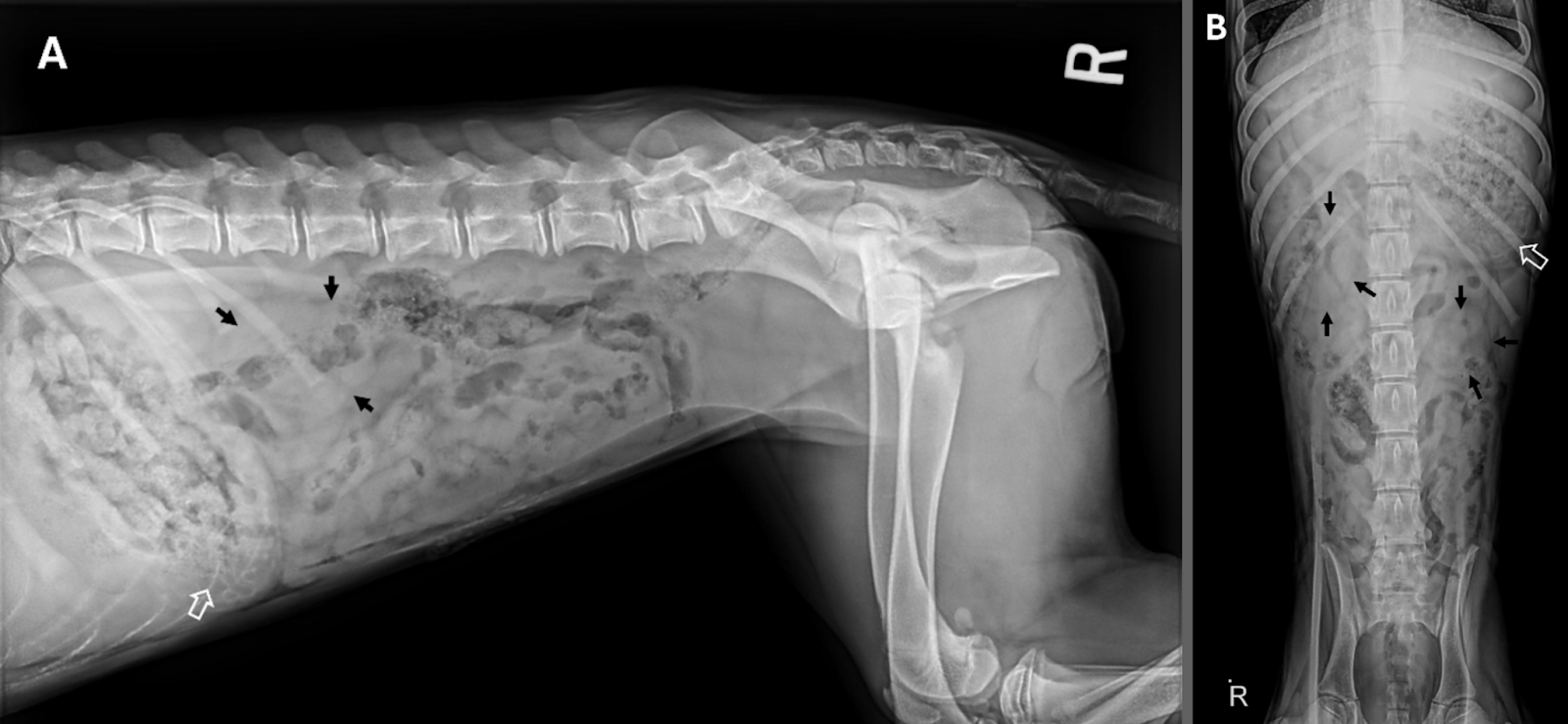
A 5 month old, female spayed, labrador cross, presented for a 3-4 week history of vomiting and hyporexia. The patient had a thin body condition on physical exam. On CBC, the patient had mild, nonregenerative, normocytic, normochromic anemia (HCT 31%) and mild lymphocytosis (4.93*10^9/L). On chemistry, the SDMA was markedly elevated (89 µg/dL) and the patient was severely azotemic (Cr 6.24 mmol/L, BUN 48 mmol/L), moderately hyperphosphotemic (5.0 mmol/L), mildly hypochloremic (103 mmol/L), mildly hypoproteinemic (5.4 dg/L while normoglobulinemic and normoalbumenemic), and mildly elevated ALP (172 IU/L). A single lateral thoracic radiograph was unremarkable. An abdominal radiograph series showed decreased peritoneal detail consistent with patient age, a moderately distended stomach with mineral opaque rugal folds, and bilaterally enlarged and irregularly margined kidneys. An abdominal ultrasound was performed and confirmed bilaterally enlarged, markedly malformed kidneys and a hyperechoic band within the gastric mucosal layer. A diagnosis of bilateral renal dysplasia with secondary uremic gastropathy was given.
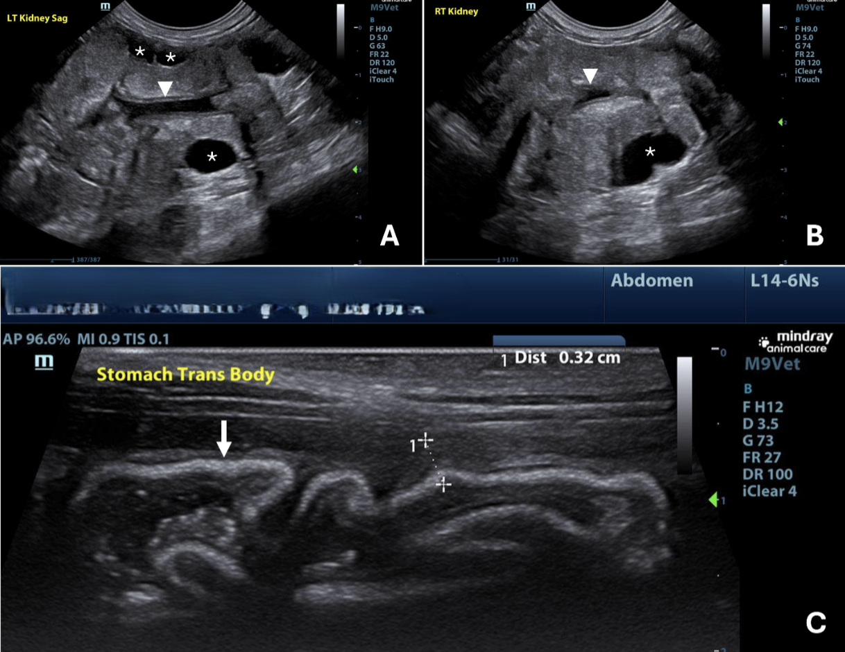
(A) and transverse image of the right kidney (B) show enlarged and malformed kidneys, indicative of renal dysplasia. The white arrow heads show compression of the renal diverticulum secondary to corticomedullary enlargement. Multiple cortical cysts are highlighted with a white asterisk. A transverse image of the stomach (C) shows gastric wall thickening (dotted bracket) and a hyperechoic, mucosal band (white arrow) consistent with metastatic mineralization. Renal dysplasia is a congenital condition characterized by disorganized development of renal parenchyma and is seen in dogs both with and without a hereditary component.
Consequently, clinically affected dogs develop chronic kidney disease (CKD) early in life. Sequelae of severely affected individuals include uremia, decreased clearance of gastrin (a hormone that stimulates gastric acid secretion), and a high calcium-phosphorus product, as seen in this case. This increased systemic urea diffuses from interstitial fluid into various tissues, including the gastric mucosa and vasculature namely in the gastric mucosa and submucosa 1 . Subsequent gastric ulceration, necrosis, and metastatic mineralization result in the pathognomonic sonographic feature of a hyperechoic band within the gastric mucosa. In such cases, treatment revolves around palliative care.
Sources
- 1. Puccinelli, Caterina et al. “Ultrasonographic Features of Gastropathy in Dogs with Acute Kidney Injury and Acute-on-Chronic Kidney Injury.” Animals : an open access journal from MDPI vol. 15,18 2666. 11 Sep. 2025,
- doi:10.3390/ani15182666
